Cancer Research

Newswise — A simple blood test can rapidly and accurately detect mutations in two key genes in non-small cell lung tumors, researchers at Dana-Farber Cancer Institute and other institutions report in a new study – demonstrating the test’s potential as a clinical tool for identifying patients who can benefit from drugs targeting those mutations. The test, known as a liquid biopsy, proved so reliable in the study that the Dana-Farber/Brigham and Women’s Cancer Center (DF/BWCC) this week became the first medical facility in the country to offer it to all patients with non-small cell lung cancer (NSCLC), either at the time of first diagnosis or of relapse following previous treatment. NSCLC is the most common form of lung cancer, diagnosed in more than 200,000 people in the United States each year, according to the American Cancer Society. An estimated 30 percent of NSCLC patients have mutations in either of the genes included in the study, and can often be treated with targeted therapies. The study is being published online today by the journal JAMA Oncology. The liquid biopsy tested in the study – technically known as rapid plasma genotyping – involves taking a test tube-full of blood, which contains free-floating DNA from cancer cells, and analyzing that DNA for mutations or other abnormalities. (When tumor cells die, their DNA spills into the bloodstream, where it’s known as cell-free DNA.) The technique, which provides a “snapshot” of key genetic irregularities in a tumor, is a common tool in research for probing the molecular make-up of different kinds of cancers. 
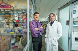
“We see plasma genotyping as having enormous potential as a clinical test, or assay – a rapid, noninvasive way of screening a cancer for common genetic fingerprints, while avoiding the challenges of traditional invasive biopsies,” said the senior author of the study, Geoffrey Oxnard, MD, thoracic oncologist and lung cancer researcher at Dana-Farber and Brigham and Women’s Hospital. “Our study was the first to demonstrate prospectively that a liquid biopsy technique can be a practical tool for making treatment decisions in cancer patients. The trial was such a success that we are transitioning the assay into a clinical test for lung cancer patients at DF/BWCC.” The study involved 180 patients with NSCLC, 120 of whom were newly diagnosed, and 60 of whom had become resistant to a previous treatment, allowing the disease to recur. Participants’ cell-free DNA was tested for mutations in the EGFR and KRAS genes, and for a separate mutation in EGFR that allows tumor cells to become resistant to front-line targeted drugs. The test was performed with a technique known as droplet digital polymerase chain reaction (ddPCR), which counts the individual letters of the genetic code in cell-free DNA to determine if specific mutations are present. Each participant also underwent a conventional tissue biopsy to test for the same mutations. The results of the liquid biopsies were then compared to those of the tissue biopsies. The data showed that liquid biopsies returned results much more quickly. The median turnaround time for liquid biopsies was three days, compared to 12 days for tissue biopsies in newly diagnosed patients and 27 days in drug-resistant patients. Liquid biopsy was also found to be highly accurate. In newly diagnosed patients, the “predictive value” of plasma ddPCR was 100 percent for the primary EGFR mutation and the KRAS mutation – meaning that a patient who tested positive for either mutation was certain to have that mutation in his or her tumor. For patients with the EGFR resistance mutation, the predictive value of the ddPCR test was 79 percent, suggesting the blood test was able to find additional cases with the mutation that were missed using standard biopsies. “In some patients with the EGFR resistance mutation, ddPCR detected mutations missed by standard tissue biopsy,” Oxnard remarked. “A resistant tumor is inherently made up of multiple subsets of cells, some of which carry different patterns of genetic mutations. A single biopsy is only analyzing a single part of the tumor, and may miss a mutation present elsewhere in the body. A liquid biopsy, in contrast, may better reflect the distribution of mutations in the tumor as a whole.” When ddPCR failed to detect these mutations, the cause was less clear-cut, Oxnard says. It could indicate that the tumor cells don’t carry the mutations or, alternatively, that the tumor isn’t shedding its DNA into the bloodstream. This discrepancy between the test results and the presence of mutations was less common in patients whose cancer had metastasized to multiple sites in the body, researchers found. The ddPCR-based test, or assay, was piloted and optimized for patients at the Translational Resarch lab of the Belfer Center for Applied Cancer Science at Dana-Farber. It was then validated for clinical use at Dana-Farber’s Lowe Center for Thoracic Oncology. An advantage of this form of liquid biopsy is that it can help doctors quickly determine whether a patient is responding to therapy. Fifty participants in the study had repeat testing done after starting treatment for their cancer. “Those whose blood tests showed a disappearance of the mutations within two weeks were more likely to stay on the treatment than patients who didn’t see such a reduction,” said the study’s lead author, Adrian Sacher, MD, of Dana-Farber and Brigham and Women’s Hospital. And because tumors are constantly evolving and acquiring additional mutations, repeated liquid biopsies can provide early detection of a new mutation – such as the EGFR resistance mutation – that can potentially be treated with targeted agents. “The study data are compelling,” said DF/BWCC pathologist Lynette Sholl, MD, explaining the center’s decision to begin offering ddPCR-based liquid biopsy to all lung cancer patients. “We validated the authors’ findings by cross-comparing results from liquid and tissue biopsies in 34 NSCLC patients. To work as a real-world clinical test, liquid biopsy needs to provide reliable, accurate data and be logistically practical. That’s what we’ve seen with the ddPCR-based blood test. “The test has great utility both for patients newly diagnosed with NSCLC and for those with a recurrence of the disease,” she continued. “It’s fast, it’s quantitative (it indicates the amount of mutant DNA in a sample), and it can be readily employed at a cancer treatment center.” The co-authors of the study are Cloud Paweletz, PhD, Allison O’Connell, BSc, and Nora Feeney, BSc, of the Belfer Center for Applied Cancer Science at Dana-Farber; Ryan S. Alden BSc, and Stacy L. Mach BA, of Dana-Farber; Suzanne E. Dahlberg, PhD, of Dana-Farber and Harvard T.H. Chan School of Public Health; and Pasi A. Jänne, MD, PhD, of Dana-Farber, the Belfer Center, and Brigham and Women’s Hospital.
Newswise — People in addiction treatment programs around the world use tobacco at two to three times the rate of people who are not being treated for addiction, according to a review of research studies from 20 countries other than the United States.  “When people come into treatment for drugs and alcohol, we are not treating another addiction that has a significant chance of eventually killing them, which is tobacco use,” said Guydish. “At a public health level, this means that our addiction treatment efforts should address smoking and tobacco use better than they do now.”  Guydish and his team reviewed 54 studies, involving a total of 37,364 participants in 20 countries on six continents, which were published in English from 1987 to 2013. They found that among people in treatment for drug and alcohol use, the overall rate of smoking was 84 percent, compared with a rate of 31 percent for members of the general population, matched for gender and year of study.  The results agree with an earlier review led by Guydish of smoking addiction treatment programs in the U.S. In that paper, the authors found that the median smoking rate among people in addiction treatment was 76.3 percent, in contrast with the smoking rate in the general U.S. population, which is now estimated at less than 18 percent. “Every person who enters substance abuse treatment ought to have their tobacco use evaluated and treated,” said Guydish. “If they don’t want to be treated and quit right away, they should have some education to help them think more about quitting.”  Guydish observed that “there are data from a number of studies which strongly suggest that you can improve substance treatment outcomes by addressing smoking among the patients in treatment. That’s what we should be doing.”  The World Health Organization (WHO) has created a policy package called MPOWER, noted Guydish, which is designed to assist countries in implementing anti-smoking initiatives. “We would recommend that WHO pay attention to this finding and use it to extend their MPOWER strategies,” said Guydish. “Anyone who is interested in smoking reduction internationally could use this information at the policy level.”  Co-authors of the study are Emma Passalacqua, Anna Pagano, PhD, Thao Le, MPH, Barbara Tajima, MEd, Lindsay Docto, Daria Garina and Kevin Delucchi, PhD, of UCSF; Cristina Martínez of the Catalan Institute of Oncology-Institut d'Investigació Biomèdica de Bellvitge, Barcelona, Spain; and JongSerl Chun of Ewha Womans University, Seoul, South Korea. The paper is titled, “An International Systematic Review of Smoking Prevalence in Addiction Treatment.” The study was supported by funds from the National Institute on Drug Abuse and the UCSF Tobacco Related Disease Research Program. UCSF is the nation's leading university exclusively focused on health. Now celebrating the 150th anniversary of its founding as a medical college, UCSF is dedicated to transforming health worldwide through advanced biomedical research, graduate-level education in the life sciences and health professions, and excellence in patient care. It includes top-ranked graduate schools of dentistry, medicine, nursing and pharmacy; a graduate division with world-renowned programs in the biological sciences, a preeminent biomedical research enterprise and top-tier hospitals, UCSF Medical Center and UCSF Benioff Children's Hospitals.    
Newswise — Jan. 22, 2016─A diet rich in fiber may not only protect against diabetes and heart disease, it may reduce the risk of developing lung disease, according to new research published online, ahead of print in the Annals of the American Thoracic Society. Analyzing data from the National Health and Nutrition Examination Surveys, researchers report in “The Relationship between Dietary Fiber Intake and Lung Function in NHANES,” that among adults in the top quartile of fiber intake: • 68.3 percent had normal lung function, compared to 50.1 percent in the bottom quartile. • 14. 8 percent had airway restriction, compared to 29.8 percent in the bottom quartile.In two important breathing tests, those with the highest fiber intake also performed significantly better than those with the lowest intake. Those in the top quartile had a greater lung capacity (FVC) and could exhale more air in one second (FEV1) than those in the lowest quartile. “Lung disease is an important public health problem, so it’s important to identify modifiable risk factors for prevention,” said lead author Corrine Hanson PhD, RD, an associate professor of medical nutrition at the University of Nebraska Medical Center. “However, beyond smoking very few preventative strategies have been identified. Increasing fiber intake may be a practical and effective way for people to have an impact on their risk of lung disease.” Researchers reviewed records of 1,921 adults, ages 40 to 79, who participated in NHANES during 2009-2010. Administered by the Centers for Disease Control and Prevention, NHANES is unique in that it combines interviews with physical examinations. Fiber consumption was calculated based on the amount of fruits, vegetables, legumes and whole grains participants recalled eating. Those whose diets included more than 17.5 grams of fiber a day were in the top quartile and represented the largest number of participants, 571. Those getting less than 10.75 grams of fiber a day were in the lower group and represented the smallest number of participants, 360. Researchers adjusted for a number of demographic and health factors, including smoking, weight and socioeconomic status, and found an independent association between fiber and lung function. They did not adjust for physical activity, nor did the NHANES data allow them to analyze fiber intake and lung function over time—limitations acknowledged by the authors. Authors cited previous research that may explain the beneficial effects of fiber they observed. Other studies have shown that fiber reduces inflammation in the body, and the authors noted that inflammation underlies many lung diseases. Other studies have also shown that fiber changes the composition of the gut microbiome, and the authors said this may in turn reduce infections and release natural lung-protective chemicals to the body. If further studies confirm the findings of this report, Hanson believes that public health campaigns may one day “target diet and fiber as safe and inexpensive ways of preventing lung disease.” To read the article in full, please visit: http://www.thoracic.org/about/newsroom/press-releases/resources/White-201509-609OC.PDF
Newswise — DURHAM, N.C. -- Doctors at the Duke University School of Medicine have tested a new injectable agent that causes cancer cells in a tumor to fluoresce, potentially increasing a surgeon’s ability to locate and remove all of a cancerous tumor on the first attempt. The imaging technology was developed through collaboration with scientists at Duke, the Massachusetts Institute of Technology (MIT) and Lumicell Inc. According to findings published January 6 in Science Translational Medicine, a trial at Duke University Medical Center in 15 patients undergoing surgery for soft-tissue sarcoma or breast cancer found that the injectable agent, a blue liquid called LUM015 (loom – fifteen), identified cancerous tissue in human patients without adverse effects. Cancer surgeons currently rely on cross-sectional imaging such as MRIs and CT scans to guide them as they remove a tumor and its surrounding tissue. But in many cases some cancerous tissue around the tumor is undetected and remains in the patient, sometimes requiring a second surgery and radiation therapy. “At the time of surgery, a pathologist can examine the tissue for cancer cells at the edge of the tumor using a microscope, but because of the size of cancer it’s impossible to review the entire surface during surgery,” said senior author David Kirsch, M.D., Ph.D., a professor of radiation oncology and pharmacology and cancer biology at Duke University School of Medicine. “The goal is to give surgeons a practical and quick technology that allows them to scan the tumor bed during surgery to look for any residual fluorescence.” Researchers around the globe are pursuing techniques to help surgeons better visualize cancer, some using a similar mechanism as LUM015, which is activated by enzymes. But the Duke trial described in the journal is the first protease-activated imaging agent for cancer that has been tested for safety in humans, Kirsch said. LUM015 was developed by Lumicell, a company started by researchers at MIT and involving Kirsch. In companion experiments in mice described in the journal, LUM015 accumulated in tumors where it creates fluorescence in tumor tissue that is on average five times brighter than regular muscle. The resulting signals aren’t visible to the naked eye and must be detected by a handheld imaging device with a sensitive camera, which Lumicell is also developing, Kirsch said. In the operating room after a tumor is removed, surgeons would place the handheld imaging device on the cut surface. The device would alert them to areas with fluorescent cancer cells. Going into surgery, the goal is always to remove 100 percent of the tumor, plus a margin of normal tissue around the edges, explained senior author Brian Brigman, M.D., Ph.D., chief of orthopedic oncology at Duke. Pathologists then analyze the margins over several days and determine whether they are clear. “This pathologic technique to determine whether tumor remains in the patient is the best system we have currently, and has been in use for decades, but it’s not as accurate as we would like,” said Brigman, who is also the director of the sarcoma program at the Duke Cancer Institute. “If this technology is successful in subsequent trials, it would significantly change our treatment of sarcoma. If we can increase the cases where 100 percent of the tumor is removed, we could prevent subsequent operations and potentially cancer recurrence. Knowing where there is residual disease can also guide radiation therapy, or even reduce how much radiation a patient will receive.” Researchers at Massachusetts General Hospital are currently evaluating the safety and efficacy of LUM015 and the Lumicell imaging device in a prospective study of 50 women with breast cancer. Afterward, Kirsch said, multiple institutions would likely evaluate whether the technology can decrease the number of patients needing subsequent operations following initial breast cancer removal. In addition to Kirsch and Brigman, study authors include Melodi Javid Whitley, Diana M. Cardona, Alexander L. Lazarides, Ivan Spasojevic, Jorge M. Ferrer, Joan Cahill, Chang-Lung Lee, Matija Snuderl, Dan G. Blazer III, E. Shelley Hwang, Rachel A. Greenup, Paul J. Mosca, Jeffrey K. Mito, Kyle C. Cuneo, Nicole A. Larrier, Erin K. O’Reilly, Richard F. Riedel, William C. Eward, David B. Strasfeld, Dai Fukumura, Rakesh K. Jain, W. David Lee, Linda G. Griffith and Moungi G. Bawendi. Duke author Kirsch and MIT authors Griffith, Bawendi, Ferrer and W. David Lee hold interest in or are involved with Lumicell Inc., a company commercializing LUM015 and the imaging system. Duke and MIT hold a patent on the imaging device technology. More detailed conflict-of-interest information is included in the manuscript published by Science Translational Medicine. The study was funded in part by an American Society of Clinical Oncology Advanced Clinical Research Award to Kirsch, the National Institutes of Health (NIH) (T32GM007171), a National Cancer Institute Small Business Innovation Research award to Lumicell Inc. (1U43CA165024), the NIH National Center for Advancing Translational Science (UL1TR001117), and Duke Comprehensive Cancer Center Support (5P30-CA-014236-38). Lumicell Inc. provided the imaging agents.
Newswise — Stony Brook, NY, Embargoed Until 1 PM, EST; December 16, 2015 – A team of researchers from Stony Brook University, led by Yusuf Hannun, MD, the Joel Strum Kenny Professor in Cancer Research and Director of the Stony Brook University Cancer Center, have found quantitative evidence proving that extrinsic risk factors, such as environmental exposures and behaviors weigh heavily on the development of a vast majority (approximately 70 to 90 percent) of cancers. The finding, reported in the December 16 online issue of Nature, in a paper titled “Substantial contribution of extrinsic risk factors to cancer development,” may be important for strategizing cancer prevention, research and public health. Inspired by a January 2015 research paper in Science, which concluded that the majority of the variation in cancer risk among tissues is due to “bad luck,” the Stony Brook team used the same data to assess what leads to the risk of developing cancer. The interdisciplinary team of researchers from the Departments of Applied Mathematics and Statistics, Medicine, Pathology and Biochemistry, concluded the opposite – that most cancers are the result of external risk factors. “Cancer is caused by mutations in the DNA of cells, which leads to uncontrolled cell growth instead of orderly growth. But the development of cancer is a complex issue, and we as a scientific community need to have solid analytical models to investigate what intrinsic and extrinsic factors cause certain forms of cancer,” said Dr. Hannun, senior author of the paper. “Many scientists argued against the ‘bad luck’ or ‘random mutation’ theory of cancer but provided no alternative analysis to quantify the contribution of external risk factors,” explained Song Wu, PhD, lead author of the paper, and Assistant Professor in the Department of Applied Mathematics and Statistics, Stony Brook University. “Our paper provides an alternative analysis by applying four distinct analytic approaches.” They developed four distinct approaches to assess cancer risk. With these four approaches, they discovered collectively and individually that most cancers are attributed largely to external risk factors, with only 10-to-30 percent attributed to random mutations, or intrinsic factors. First, the researchers examined extrinsic risks by tissue cell turnover. In a data-driven approach, they re-examined the quantitative relationship between observed lifetime risk of cancer (ie, for lung, pancreatic, colorectal and other tissues) and division of the normal tissue stem cells in those groups reported in the Science paper. If intrinsic risk factors played a major role, the tissue with the similar stem cell divisions would show similar observed lifetime cancer risk. They found this pattern to be a rare one, and thus determined intrinsic factors played a vital role in only about 10 percent of cancers. These results are supported by strong epidemiologic evidence; for example studies showing that immigrants moving from countries with lower cancer incidence to countries with higher rates of cancer incidence acquire the higher risk in their new country. The researchers also mathematically surveyed and analyzed recent studies on mutational signatures in cancer, which are regarded as “fingerprints” left on cancer genomes by different mutagenic processes. Some 30 distinct signatures among various cancers were identified. They analyzed the signatures and categorized them as having intrinsic or extrinsic origins. They found that while a few forms of cancer had a greater than 50 percent of intrinsic mutations, the majority of cancers, such as colorectal, lung, bladder and thyroid cancers had large proportions of mutations likely caused by extrinsic factors. The team also analyzed the SEER (Surveillance, Epidemiologic and End Results Program) data, which showed that many cancers have been increasing in incidence and in mortality, suggesting that external factors contribute heavily to these cancers. Lastly, they used computational modeling to dissect the contribution of the intrinsic processes in the development of cancer, based on known gene mutations in cancer and the likelihood that they arise from intrinsic mutation rates. They found that when three or more mutations are required for cancer onset (which is a currently accepted parameter), intrinsic factors are far from sufficient to account for the observed risks, indicating small percentages of intrinsic cancer risks in many cancers. The four methods involved both data- and model-driven quantitative analyses, with and without using the stem cell estimations. The idea behind the overall approach was to assess cancer risk by multiple methods and not by a single type of analysis. Dr. Hannun concluded that their overall approach “provides a new framework to quantify the lifetime cancer risks from both intrinsic and extrinsic factors, which will have important consequences for strategizing cancer prevention, research and public health.” Co-authors of the paper include: Scott Powers of the Department of Pathology at Stony Brook University, and Wei Zhu, of the Department of Applied Mathematics and Statistics at Stony Brook University. All of the authors are collaborating investigators at the Stony Brook University Cancer Center. ###About Stony Brook University Part of the State University of New York system, Stony Brook University encompasses 200 buildings on 1,450 acres. Since welcoming its first incoming class in 1957, the University has grown tremendously, now with more than 25,000 students and 2,500 faculty. Its membership in the prestigious Association of American Universities (AAU) places Stony Brook among the top 62 research institutions in North America. U.S. News & World Report ranks Stony Brook among the top 100 universities in the nation and top 40 public universities, and Kiplinger names it one of the 35 best values in public colleges. One of four University Center campuses in the SUNY system, Stony Brook co-manages Brookhaven National Laboratory, putting it in an elite group of universities that run federal research and development laboratories. A global ranking by U.S. News & World Report places Stony Brook in the top 1 percent of institutions worldwide. It is one of only 10 universities nationwide recognized by the National Science Foundation for combining research with undergraduate education. As the largest single-site employer on Long Island, Stony Brook is a driving force of the regional economy, with an annual economic impact of $4.65 billion, generating nearly 60,000 jobs, and accounts for nearly 4 percent of all economic activity in Nassau and Suffolk counties, and roughly 7.5 percent of total jobs in Suffolk County. Greg FilianoMedia Relations ManagerSchool of MedicineStony Brook UniversityOffice of Communications and Marketing631-444-9343Gregory.filiano@stonybrookmedicine.edu
Newswise — There are two common approaches to protecting humans from infectious disease: Targeting pathogens and parasites with medicines like antibiotics, or dealing with the conditions that allow transmission. A paper published today in the journal Nature Scientific Reports demonstrates the effectiveness of a third strategy: Adjusting the landscape of the human body to remove the mechanism that allows pathogens to cause disease. The discovery is the result of serendipity and collaboration between high-level scientists in different fields. "It was pure luck that I ended up on this paper," says Dan Theodorescu, MD, PhD, director of the University of Colorado Cancer Center. "Bill Petri and I had been social friends for years – Christmas parties, that kind of thing. When I was at Virginia it happened that we were on a recruitment committee together and the candidate was late, so we started talking." His conversation with William A. Petri, Jr., MD, PhD, chief of the Division of Infectious Diseases & International Health at the University of Virginia led to the idea of applying an innovative cancer science technique to the study of infectious disease. With first author Chelsea Marie, PhD, postdoctoral researcher in the Petri Laboratory at Virginia, the group decided to silence genes in human cells to discover if the loss of any single gene would confer immunity to the parasite E. histolytica, which infects 50 million people and causes 40,000-110,000 deaths via severe diarrhea worldwide. "Chelsea is a fearless experimenter. She took a library of cells that Dan had developed in his work with bladder cancer and then sequentially killed them with E. histolytica parasites," Petri says. Specifically, the group used the technique called RNAi to create a library of bladder cancer cells with thousands of independent, silenced genes. Then they challenged these cultures with the parasite E. histolytica. "We do this all the time in cancer research," Theodorescu says. "Commonly, we're looking for genes that, when silenced, will make cells more susceptible to chemotherapy." In this case the analogue of chemotherapy was the infectious, dangerous pathogen. "This amoeba is a cluster bomb – a voracious killer. In the back of my mind I was thinking the parasite was going to decimate the host cells no matter what we did with their genetics," Marie says. For the vast majority of cells in this genome-wide screen, Chelsea Marie was correct; E. histolytica decimated many thousands of these independent cell cultures. However, a small number of cells seemed to resist the parasite. Was this the random chance of lucky survival or had silenced genes somehow offered immunity to these cells? To find out, Marie discarded the killed cells and retested the cells that had survived; again she infected these survivor cells with E. histolytica. "It wasn't a fluke," says Marie. "We did this over nine generations of cells, each time selecting the cells that survived and then re-applying the parasite. Over these generations of selection, we saw the cultures becoming more and more enriched for cells lacking specific genes." Using next generation sequencing, Marie identified the genes that conferred resistance and found that many were involved in managing the flow of potassium into and out of human cells. Specifically, the identified genes KCNA3, KCNB2, KCNIP4, KCNJ3, and SLC24A3 are involved in what is called potassium transport. A follow-up experiment showed that new intestinal cells treated with E. histolytica showed potassium efflux – the flow of potassium from inside a cell out through the cell wall – directly before cell death. "We started to see a pretty clear line of reasoning," says Theodorescu. "The parasite was causing potassium efflux right before cell death and cells that happened to be unable to transport potassium didn't die." To ensure that lack of potassium transport was, in fact, causing resistance to the parasite, the group reversed the direction of their experiments. Marie started with new cells and used drugs to block their ability to transport potassium. Blocking potassium efflux created cells that were resistant to E. histolytica. "There is a clear need for new drugs targeting E. histolytica," Petri says. "Right now there is a single antibiotic that works against this parasite. We know that eventually the parasite will develop resistance to the antibiotic and at that point there's no plan B. This could be the plan B – targeting the human genes that enable the parasite to cause disease." Marie is pushing forward. She recently learned from a mentor at John's Hopkins how to isolate stem cells from human tissue to grow what she calls "mini guts" to test therapeutics that may be useful in human patients. And technological advances make this study's general technique more efficient, allowing the use of what are called CRISPR libraries instead of RNAi screens. "This is a major finding with translational implications for this infection that causes so many deaths worldwide, but also proof that this cancer-science approach can be used to explore genetic mechanisms of resistance in the field of infectious disease," Theodorescu says. The field of infectious disease has been focused on the infection, targeting pathogens and their transmission. This study shows that in addition to characteristics of the parasite, mortality due to disease can be prevented by manipulating characteristics of the host. 
Newswise — New Brunswick, N.J., September 8, 2015– Rutgers Cancer Institute of New Jersey is one of a few East Coast sites to offer a clinical trial investigating an experimental drug known as REGN1979 in the treatment of non-Hodgkin’s lymphoma (NHL) and chronic lymphocytic leukemia (CLL). The drug – designed to use the body’s own defenses to fight illness – targets a specific protein (called CD20) found in these particular types of cancer and targets another protein (called CD3) found on T-cells, a type of cell in the immune system. REGN1979 is designed to help T cells find and destroy B cells, including those cancerous B cells found in NHL and CLL. The goal is to determine how much of the drug can be given safely to patients who have the CD20 protein on their lymphoma or CLL cells. “By harnessing the body’s own natural defenses, there is an opportunity to provide alternate therapies for patients with NHL and CLL whose disease has stopped responding to standard treatments,” notes Rajat Bannerji, MD, PhD, medical oncologist and principal investigator of the trial at Rutgers Cancer Institute of New Jersey and associate professor of medicine at Rutgers Robert Wood Johnson Medical School. Participants are expected to be involved in the study for at least one year. Patients enrolled into the study will receive an infusion of the study drug through a vein. Participants also will be asked permission for scientists to study tissue samples taken from tumors or bone marrow collected during certain clinic visits. Adults aged 18 and older who are diagnosed with NHL or CLL and have had prior treatment with a particular antibody therapy (anti-CD20) are eligible to take part in the trial, provided they meet additional entry criteria. Prior to being enrolled into the study, participants would be required to undergo a number of tests including blood work and a physical exam. For more information on how to take part in this trial, sponsored by Regeneron Pharmaceuticals, Inc., individuals should call the Cancer Institute’s Office of Human Research Services at 732-235-8675 or e-mail cinjclinicaltrials@cinj.rutgers.edu. Clinical trials, often called cancer research studies, test new treatments and new ways of using existing treatments for cancer. At the Cancer Institute, researchers use these studies to answer questions about how a treatment affects the human body and to make sure it is safe and effective. There are several types of clinical trials that are currently underway at the Cancer Institute, including those that diagnose, treat, prevent, and manage symptoms of cancer. Many treatments used today, whether they are drugs or vaccines, ways to do surgery or give radiation therapy, or combinations of treatments, are the results of past clinical trials. As New Jersey’s only National Cancer Institute-designated Comprehensive Cancer Center, the Cancer Institute offers patients access to treatment options not available at other institutions within the state. The Cancer Institute currently enrolls more than 1,200 patients in clinical trials annually, including approximately 17 percent of all new adult cancer patients and approximately 70 percent of all pediatric cancer patients. Enrollment in these studies nationwide is fewer than five percent of all adult cancer patients. About Rutgers Cancer Institute of New JerseyRutgers Cancer Institute of New Jersey (www.cinj.org) is the state’s first and only National Cancer Institute-designated Comprehensive Cancer Center. As part of Rutgers, The State University of New Jersey, the Cancer Institute of New Jersey is dedicated to improving the detection, treatment and care of patients with cancer, and to serving as an education resource for cancer prevention. Physician-scientists at the Cancer Institute engage in translational research, transforming their laboratory discoveries into clinical practice, quite literally bringing research to life. To make a tax-deductible gift to support the Cancer Institute of New Jersey, call 848-932-3637 or visit www.cinj.org/giving. Follow us on Facebook at www.facebook.com/TheCINJ. The Cancer Institute of New Jersey Network is comprised of hospitals throughout the state and provides the highest quality cancer care and rapid dissemination of important discoveries into the community. Flagship Hospital: Robert Wood Johnson University Hospital. System Partner: Meridian Health (Jersey Shore University Medical Center, Ocean Medical Center, Riverview Medical Center, Southern Ocean Medical Center, and Bayshore Community Hospital). Major Clinical Research Affiliate Hospitals: Carol G. Simon Cancer Center at Morristown Medical Center and Carol G. Simon Cancer Center at Overlook Medical Center. Affiliate Hospitals: JFK Medical Center, Robert Wood Johnson University Hospital Hamilton (CINJ Hamilton), and Robert Wood Johnson University Hospital Somerset.
Newswise — ST. LOUIS — In research published inCancer Cell, Thomas Burris, Ph.D., chair of pharmacology and physiology at Saint Louis University, has, for the first time, found a way to stop cancer cell growth by targeting the Warburg Effect, a trait of cancer cell metabolism that scientists have been eager to exploit. Unlike recent advances in personalized medicine that focus on specific genetic mutations associated with different types of cancer, this research targets a broad principle that applies to almost every kind of cancer: its energy source. The Saint Louis University study, which was conducted in animal models and in human tumor cells in the lab, showed that a drug developed by Burris and colleagues at Scripps Research Institute can stop cancer cells without causing damage to healthy cells or leading to other severe side effects. The Warburg EffectMetabolism – the ability to use energy – is a feature of all living things. Cancer cells aggressively ramp up this process, allowing mutated cells to grow unchecked at the expense of surrounding tissue. “Targeting cancer metabolism has become a hot area over the past few years, though the idea is not new,” Burris said. Since the early 1900s, scientists have known that cancer cells prefer to use glucose as fuel even if they have plenty of other resources available. In fact, this is how doctors use PET (positron emission tomography) scan images to spot tumors. PET scans highlight the glucose that cancer cells have accumulated. This preference for using glucose as fuel is called the Warburg effect, or glycolysis. In his paper, Burris reports that the Warburg effect is the metabolic foundation of oncogenic (cancer gene) growth, tumor progression and metastasis as well as tumor resistance to treatment. Cancer’s Goal: To Grow and DivideCancer cells have one goal: to grow and divide as quickly as possible. And, while there are a number of possible molecular pathways a cell could use to find food, cancer cells have a set of preferred pathways. “In fact, they are addicted to certain pathways,” Burris said. “They need tools to grow fast and that means they need to have all of the parts for new cells and they need new energy.” “Cancer cells look for metabolic pathways to find the parts to grow and divide. If they don’t have the parts, they just die,” said Burris. “The Warburg effect ramps up energy use in the form of glucose to make chemicals required for rapid growth and cancer cells also ramp up another process, lipogenesis, that lets them make their own fats that they need to rapidly grow.” If the Warburg effect and lipogenesis are key metabolic pathways that drive cancer progression, growth, survival, immune evasion, resistance to treatment and disease recurrence, then, Burris hypothesizes, targeting glycolysis and lipogenesis could offer a way to stop a broad range of cancers. Cutting off the Energy SupplyBurris and his colleagues created a class of compounds that affect a receptor that regulates fat synthesis. The new compound, SR9243, which started as an anti-cholesterol drug candidate, turns down fat synthesis so that cells can’t produce their own fat. This also impacts the Warburg pathway, turning cancer cells into more normal cells. SR9243 suppresses abnormal glucose consumption and cuts off cancer cells’ energy supply. When cancer cells don’t get the parts they need to reproduce through glucose or fat, they simply die. Because the Warburg effect is not a feature of normal cells and because most normal cells can acquire fat from outside, SR9243 only kills cancer cells and remains non-toxic to healthy cells. The drug also has a good safety profile; it is effective without causing weight loss, liver toxicity, or inflammation. Promising ResultsSo far, SR9243 has been tested in cultured cancer cells and in human tumor cells grown in animal models. Because the Warburg pathway is a feature of almost every kind of cancer, researchers are testing it on a number of different cancer models. “It works in a wide range of cancers both in culture and in human tumors developing in animal models,” Burris said. “Some are more sensitive to it than others. In several of these pathways, cells had been reprogramed by cancer to support cancer cell growth. This returns the metabolism to that of more normal cells.” In human tumors grown in animal models, Burris said, “It worked very well on lung, prostate, and colorectal cancers, and it worked to a lesser degree in ovarian and pancreatic cancers.” It also seems to work on glioblastoma, an extremely difficult to treat form of brain cancer, though it isn’t able to cross the brain/blood barrier very effectively. The challenge for researchers in this scenario will be to find a way to allow the drug to cross this barrier, the body’s natural protection for the brain, which can make it difficult for drug treatments to reach their target. And, in even more promising news, it appears that when SR9243 is used in combination with existing chemotherapy drugs, it increases their effectiveness, in a mechanism apart from SR9243’s own cancer fighting ability. Other researchers on the study include Colin A. Flaveny, Kristine Griffett, Bahaa El-Dien M. El-Gendy, Melissa Kazantzis, Monideepa Sengupta, Antonio L. Amelio, Arindam Chatterjee, John Walker, Laura A. Solt and Theodore M. Kamenecka. Established in 1836, Saint Louis University School of Medicine has the distinction of awarding the first medical degree west of the Mississippi River. The school educates physicians and biomedical scientists, conducts medical research, and provides health care on a local, national and international level. Research at the school seeks new cures and treatments in five key areas: cancer, liver disease, heart/lung disease, aging and brain disease, and infectious diseases.