News

Newswise —
Obese young people can still turn their chances of developing life threatening illness around if they change before middle age, says new research.
The study looked at the body mass index (BMI) of people when they were young and compared it to when they were middle aged to see whether it affected their risk of heart attack, stroke or diabetes.
Men who had high BMI levels at 21, but had lowered their BMI by the time they were 50, had similar or lower rates of diabetes as people who were normal weight when younger, the results showed.
In a unique approach, the study used the records of men’s military service, which recorded their BMI at 21, as well as participant recall and followed up with them 30 years later.
Lead research Professor Christopher Owen from St George’s University of London said the effects of high BMI early in life may be reversible.
“Even in men who carried out UK National Service and were relatively thin in early life compared to more recent men, higher levels of fatness in early adult life appear to be associated with later diabetes,” he said.
“However, effects of early body mass appear to be reversible by subsequent weight loss. These findings have important implications for Type 2 diabetes prevention, especially in more recent adults with high levels of obesity.”
But the study, which examined almost 5000 men, found that a higher BMI earlier in life did not impact on the risk of heart attack or stroke.
However, men who were obese when they were 50 had increased chances of suffering a heart attack, stroke or diabetes.
Obesity is the biggest risk factor for type 2 diabetes and over 4 million people in the UK are at high risk of developing the condition.

Newswise —
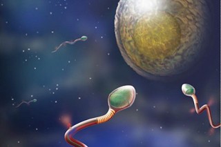
UC Berkeley biologists have discovered the switch that triggers the power kick sperm use to penetrate and fertilize a human egg, uncovering a possible source of male infertility but also a potential target for contraceptives that work in both men and women.
The switch is a protein receptor that responds to the female sex hormone progesterone, which is released by the egg or oocyte, the ultimate goal toward which sperm swim. Thousands of these receptors sit on the surface of a sperm’s tail and when the sperm gets close to the egg, the hormone activates the receptor and triggers a cascade of changes that make the tail snap like a whip, powering the sperm into and hopefully through the cells protecting the egg.
“If the receptor protein doesn’t recognize progesterone, you would be infertile,” said Melissa Miller, a postdoctoral fellow at both UC Berkeley and UC San Francisco and the first author of a paper reporting the discovery. “This gives us an understanding of another pathway that is involved in human sperm activity.”
A drug that inactivates this newly discovered receptor, however, might make a good “unisex” contraceptive – one that could be used by either sexual partner.
“What’s really cool is that we have an actual target for unisex contraceptive development,” Miller said. “If you can stop progesterone from inducing a power stroke, sperm are not going to be able to reach or penetrate the oocyte.”
While there are other possible targets for a contraceptive that would prevent the initiation of the power stroke, called hyperactivation, or the simultaneous release of enzymes that cut through the protective layer around the egg, “this is one of the better options we have for a unisex contraceptive,” she said.
Senior author Polina Lishko, a UC Berkeley assistant professor of molecular and cell biology, noted that many tissues – the brain, the lungs, smooth muscle – contain related progesterone or steroid receptors that may work in a similar manner to trigger major changes in tissues.
“Now that we know the players, the next step is to look in other tissues that express these proteins to see whether progesterone acts on them in a similar manner to affect pain threshold adjustment in pain sensing neurons, surfactant production in the lungs or the excessive smooth muscle contractions found in asthma,” she said. “This may be a universal pathway in all cells.”
Miller, Lishko and their colleagues will publish their findings in the March 17 “Fast Release” issue of the journalScience.
Few known causes of male infertility
Today, doctors are unable to determine the cause of nearly 80 percent of all cases of male infertility, in part because little is known about the many molecular steps involved in the production of sperm and its interactions with the egg. Sperm may be to blame in half of all cases of infertile couples.
Yet because the U.S. government forbids the use of federal funds for research that brings eggs and sperm together in the same dish, little research has been done on how egg-sperm interactions lead to infertility. And until five years ago, it was very difficult to study the inner workings of sperm – the body’s smallest cell – with ordinary lab techniques.
The new discovery comes thanks to techniques that Lishko and her colleague Yuriy Kirichok developed over the past five years at UCSF and UC Berkeley. The techniques allow them to stick electrodes on a sperm’s tail and record its reactions to hormones, key to probing the molecular cascades that govern sperm behavior.
That technique led to their discovery that a large receptor on sperm tails – a calcium channel dubbed CatSper – is activated by progesterone from the egg. Progesterone unlocks the channel gate, letting electrically charged calcium atoms flood into the cell. This leads to a biochemical cascade that readies the sperm cell for its last-ditch effort to fertilize the oocyte.
Miller and Lishko suspected, however, that progesterone was not acting directly on the calcium channel, but on some other receptor that, in turn, activated the calcium channel.
That proved to be the case. They showed that progesterone actually binds to a previously mysterious enzyme called ABHD2, which is found at high levels in sperm. Once progesterone binds to the enzyme, which sits on the surface of the sperm, it removes a lipid (2AG) that has been inhibiting the calcium channel. Released of inhibition, CatSper opens the gate to calcium ions and eventual sperm activation.
The inhibitor of the calcium channel CatSper is probably there for a good reason: to prevent sperm from prematurely sprinting toward the egg and using up their limited supply of energy, Miller said.
Marathon or team sport?
“People tend to think of fertilization as like a marathon, where the fastest, most powerful sperm is going to win,” Miller said. “We think of it like the Tour de France, where the riders in front are blocking the wind for the actual winner. Fertilization is a team sport, where the first sperm clear the way, expending their energy to break through the barrier cells, so that the slow and steady guy can get into the oocyte.”
The study also sheds light on a long-standing mystery about steroids like progesterone: why they appear to act in two distinctly different ways. As a sex hormone, progesterone usually triggers a cascade of events in the cell that alter the expression of genes in the nucleus, a process that can take days. But sometimes progesterone causes immediate changes in the cell – something called non-genomic steroid signaling – that evidently use a quicker process than gene expression.
Lishko, who studies the sperm of rats, mice, bulls and boars as well as human to understand how fertilization works across different species, says that sperm are a perfect system in which to study non-genomic steroid signaling, since the genes in sperm are silenced and the normal type of steroid signaling is not present. As she and her colleagues uncover the basics of steroid signaling in sperm, the same process can then be studied in many other types of cells, she said.
The research was supported by the National Institutes of Health and by Pew Scholars and Alfred P. Sloan Awards to Lishko. Lishko, Miller and Kirichok have filed a patent on usage of ABHD2.
Aside from Miller, Lishko and Kirichok, other authors of the paper are Nadja Mannowetz, Anthony Iavarone, Rojin Safavi, Rose Hill and Diana Bautista of UC Berkeley, Elena Gracheva of Yale University and James Smith of UCSF.
Healthy sperm employ a regular sinusoidal tail motion when swimming, unlike the whiplike motion they use when they reach the egg.

Newswise —
A University of California, Irvine scientific team led by infectious diseases researchers Philip Felgner and Aaron Esser-Kahn has received $8 million from the U.S. Department of Defense’s Defense Threat Reduction Agency to help develop a new vaccine for Q fever.
Caused by the Coxiella burnetii bacterium, Q fever is a highly infectious agent common among livestock. It has a history of being aerosolized for use in biological warfare and is considered a potential bioterrorism weapon.
Q fever is also a public health threat; a 2007-10 outbreak in the Netherlands affected thousands of people. Symptoms include high fever, nausea, severe headache and abdominal pain. It is rarely fatal.
“The current vaccine for Q fever is effective but has severe side effects that limit its widespread use,” said Felgner, an adjunct professor of medicine at UCI. “It’s a high priority that this vaccine be administered to members of the armed forces. Consequently, the military is interested in developing an alternative protective vaccine that’s safer and does not cause adverse reactions.”
Felgner will use an approach he pioneered at UCI to create whole proteome microarrays to discover immune response-activating antigen proteins that may be effective as a vaccine. Additionally, he’ll collaborate with Esser-Kahn, assistant professor of chemistry, whose group will develop synthetic agents that can boost and control the immune response to these proteins.
Felgner said this dual method may be applicable in creating more vaccines important to the military and general public health, adding that this is an opportunity for the Department of Defense to test these methods for their potential use against other infectious diseases.
After identifying the target proteins, Felgner will work with the U.S. Army Medical Research Institute of Infectious Diseases at Fort Detrick in Maryland on next-stage animal studies of a candidate vaccine.
The project is a successful outgrowth of the Pacific Southwest Regional Center of Excellence, one of only 11 National Institutes of Health-funded research sites dedicated to countering threats from bioterrorism agents and emerging infectious diseases. UCI received $85 million for this effort, which was led by Dr. Alan Barbour, professor of microbiology & molecular genetics. The federal program ended in 2015.
Credit Newswise —
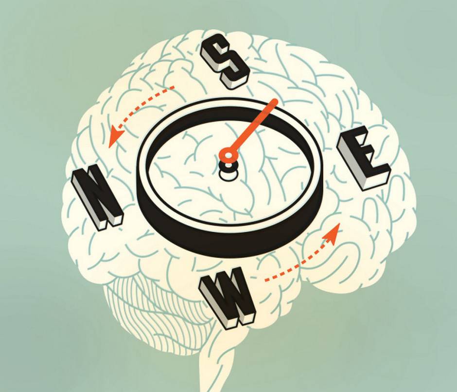
UCLA researchers have found that space-mapping neurons – the GPS system in the brain - have a strong dependence on what is being looked at when triangulating location, a finding that resolves a neurological mystery that has vexed scientists for more than four decades.
This also expands on an earlier finding that neurons responsible for creating spatial maps react differently in virtual reality than they do in the real-world environments. Researchers again used rats in a virtual reality environment to test the long-debated theory of whether landmarks are necessary or whether that region of the brain is also counting steps or directional movement to determine location, said Mayank Mehta, a UCLA professor of neurology, physics and astronomy, and neurobiology in the UCLA College and the study’s senior author.
The study, which appears today in the peer-reviewed journal Cell, showed that many neurons were firing selectively only when rats were looking at certain landmarks on screens, either in the real or in the virtual reality environment.
“This part of the brain, the hippocampus, has neurons that fire in specific places. If I’m walking around a room, some neurons fire near the door, others around the middle of the room, and they all form a map of space in the brain,” said Mehta, who also is director of a W.M. Keck Foundation neurophysics center. “Where does this map come from? The classic idea was there are two possible mechanisms. One hypothesis is that neurons triangulate distances with respect to visual landmarks. However, it was commonly believed that hippocampal neural responses do not depend on which landmark the rat was looking at, a long standing paradox. The other idea is that neurons are keeping track of the distances traveled by the subject, through the so called path-integration, though not directly tested.”
Surprisingly, the team found that the neurons did signal for what landmark the rat was looking at, thus removing the 45 year old paradox about whether the landmarks exert a causal influence on hippocampal directional responses. Further, careful experiments using virtual reality showed that the neural responses were neither abstract representations of space, as commonly thought, nor vestibular input driven path-integration signals. Instead, these responses were causally and predictably governed by visual landmarks.
This study is a part of series of studies undertaken by the Mehta laboratory to understand how the brain rapidly makes memories, including spatial maps, on the fly. They have been using Virtual Reality to manipulate the perception of space and time. Using this technique they have recently shown that in more than half of the neurons in hippocampus shut down in virtual reality. They also showed that the brain in the virtual world does not create a spatial map like it does in the real world environment, a finding that was replicated in this study, that could have implications for people who use virtual reality for gaming, military, commercial, scientific or other purposes.The scientists were studying the hippocampus, a region of the brain that is critical for learning and memory and involved in diseases such as Alzheimer’s, stroke, schizophrenia, depression, epilepsy and post-traumatic stress disorder. The hippocampus also plays an important role in forming new memories and creating mental maps of space.
Researchers created a sophisticated virtual reality environment for rats that cost nearly $1 million to develop. They placed a small harness around rats and put them on a treadmill-like device surrounded by a “virtual world” on large video screens in an otherwise dark, quiet room.
The rats walk in the virtual room in a similar way as they would in a real room, Mehta said.
Because no mental map was being made, part of the brain was not working in virtual reality. Could there be long-term implications for that as more and more people use virtual reality?
“This could have important implications. This part of the brain that makes maps of space is also involved in making memories, like what did I have for lunch, what was I doing on 9/11? It is, in that sense, what makes us human,” Mehta said. “If people are spending time in virtual reality, could that alter the way the brain works? If it’s not making maps, will the making of memories be affected? Everything in the brain influences what the brain will do later on. It’s the reason we are different at age 40 than we were at age four. The brain remembered everything that happened and modified itself because of it. Now what will happen when people’s neural responses become different in virtual reality?”Mehta said the findings, although found in rodents, are equally applicable to humans.

Credit Newswise —
A physician-researcher at The Children’s Hospital of Philadelphia (CHOP) has received a $1 million Hyundai Quantum Grant from Hyundai Hope on Wheels to advance treatment for a high-risk form of childhood leukemia. The research focuses on immunotherapy—an approach that utilizes a patient’s immune system to better fight off cancer.
“This Hyundai Quantum Grant gives us substantial resources to make progress against the most life-threatening form of leukemia,” said CHOP pediatric oncologist Richard Aplenc, M.D., Ph.D. “I’m constantly inspired by the resilience of my patients, and the grace and tenderness of their families, and this grant support is very helpful in advancing new cancer treatments.”
The Hyundai Quantum Grant, a new category in Hyundai’s pediatric cancer grant program, provides each research center $250,000 per year for four years. Focusing on pediatric cancers with the lowest survival rates, the program announced competitive awards to CHOP and three other institutions participating in the Children’s Oncology Group. Hyundai Hope on Wheels is the nonprofit organization of Hyundai Motor America, and is in its 18th year of supporting childhood cancer research. This year’s awards are Hyundai’s largest individual research grants ever for pediatric cancer research.
“We are proud to fund Dr. Aplenc, one of this year’s Hyundai Quantum winners, and his innovative work in AML,” said Dave Zuchowski, president and CEO, Hyundai Motor America. “We are hopeful for the discoveries to come from his important work in the coming years. No child should ever have to hear the words, ‘You have cancer,’ and we will continue to support the efforts fighting for that day to come.”
Overall survival rates for children’s cancer have reached 80 percent, but some high-risk forms of cancer have stubbornly resisted this progress. One such example isacute myeloid leukemia (AML), which has a complicated variety of difficult-to-treat subtypes. Even among survivors of AML, intensive chemotherapy may cause lifelong side effects.
Immunotherapy, which is currently being studied in other types of leukemia, may provide a more effective treatment for AML, with fewer long-term side effects. The new grant, said Aplenc, will enable his team to identify specific proteins on the outside surface of AML cells that could be the most appropriate targets for immune cells programmed to attack cancers.
“New technologies have dramatically altered the research landscape, by allowing scientists to better identify proteins and to make use of DNA sequencing data from AML patients,” said Aplenc. “This research grant will enable us to discover more about these specific biological molecules, offering data with great potential to help us design innovative treatments.”
Stephen Hunger, M.D., chief of the Division of Oncology and director of the Center for Childhood Cancer Researchat CHOP, added, “This award will help CHOP physicians and their collaborators develop new therapies for children with AML that has not responded to current therapies, or has relapsed despite those therapies.”
About The Children’s Hospital of Philadelphia: The Children’s Hospital of Philadelphia was founded in 1855 as the nation’s first pediatric hospital. Through its long-standing commitment to providing exceptional patient care, training new generations of pediatric healthcare professionals and pioneering major research initiatives, Children’s Hospital has fostered many discoveries that have benefited children worldwide. Its pediatric research program is among the largest in the country. In addition, its unique family-centered care and public service programs have brought the 535-bed hospital recognition as a leading advocate for children and adolescents. For more information, visit www.chop.edu.

Credit Newswise —
Could there be a vegetarian gene?
Cornell University researchers have found evidence of a genetic variation – called an allele – that has evolved in populations that have historically favored vegetarian diets, such as in India, Africa and parts of East Asia. They also discovered a different version of this gene adapted to a marine diet discovered among the Inuit in Greenland, who mainly consume seafood.
The vegetarian allele evolved in populations that have eaten a plant-based diet over hundreds of generations. The adaptation allows these people to efficiently process omega-3 and omega-6 fatty acids and convert them into compounds essential for early brain development and if they stray from a balanced omega-6 to omega-3 diet, it may make people more susceptible to inflammation, and by association, increased risk of heart disease and colon cancer.
In Inuit populations of Greenland, the researchers uncovered that a previously identified adaptation is opposite to the one found in long-standing vegetarian populations: While the vegetarian allele has an insertion of 22 bases (a base is a building block of DNA) within the gene, this insertion was found to be deleted in the seafood allele.
“The opposite allele is likely driving adaptation in Inuit,” said Kaixiong Ye, co-lead author of the paper appearing March 29 in the journal Molecular Biology and Evolution. Ye is a postdoctoral researcher in the lab of Alon Keinan, associate professor of biological statistics and computational biology, and the paper’s co-senior author.
“Our study is the first to connect an insertion allele with vegetarian diets, and the deletion allele with a marine diet,” Ye said.
“It is the most interesting example of local adaptation that I have been fortunate to help study,” said Keinan. “Several studies have pointed to adaptation in this region of the genome. Our analyses combine to show that the adaptation is driven by an insertion of a small piece of DNA that we know its function. Moreover, when it reached the Greenlandic Inuit, with their marine-based diet rich in omega-3, it might have become detrimental.”
FADS1 and FADS2 are enzymes that are essential for converting omega-3 and omega-6 fatty acids into downstream products needed for brain development and controlling inflammation. Meat and seafood eaters have less need for increased FADS1 and FADS2 enzymes to get proper nutrition because their omega-3 and omega-6 fatty acid conversion process is simpler and requires fewer steps.
This study is based on previous work by co-senior author Tom Brenna, professor of human nutrition and of chemistry at Cornell University, who showed the insertion can regulate the expression of FADS1 and FADS2 and hypothesized it could be an adaptation in vegetarian populations.
Ye, Keinan and colleagues analyzed frequencies of the vegetarian allele in 234 primarily vegetarian Indians and 311 U.S. individuals and found the vegetarian allele in 68 percent of the Indians and in just 18 percent of Americans. Analysis using data from the 1,000 Genomes Project similarly found the vegetarian allele in 70 percent of South Asians, 53 percent of Africans, 29 percent of East Asians and 17 percent of Europeans.
“Northern Europeans have a long history of drinking milk and they absorbed enough end products from milk for long-chain fatty acid metabolism so they don’t have to increase capacity to synthesize those fatty acids from precursors,” said Ye.
“One implication from our study is that we can use this genomic information to try to tailor our diet so it is matched to our genome, which is called personalized nutrition,” he added.
The researchers are not sure yet when the adaptation first occurred, as analyses of chimpanzee or orangutan genomes did not uncover the vegetarian allele. But there is evidence for the allele in early hominid Neanderthal and Denisovan genomes.
“It is possible that in the history of human evolution, when people migrated to different environments, sometimes they ate a plant-based diet and sometimes they ate a marine-based diet, and in different time periods these different alleles were adaptive,” meaning the alleles have a tendency to evolve under dietary pressures, Ye said.

Credit Newswise —
Ever see something that isn't really there? Could your mind be playing tricks on you? The "tricks" might be your brain reacting to feedback between neurons in different parts of the visual system, according to a study published in the Journal of Neuroscience by Carnegie Mellon University Assistant Professor of Biological Sciences Sandra J. Kuhlman and colleagues.
Understanding this feedback system could provide new insight into the visual system's neuronal circuitry and could have further implications for understanding how the brain interprets and understands sensory stimuli.
Many optical illusions make you see something that's not there. Take the Kanizsa triangle: when you place three Pac-Man-like wedges in the right spot, you see a triangle, even though the edges of the triangle aren't drawn.
"We see with both our brain and our eyes. Your brain is making inferences that allow you to see the triangle. It's connecting the dots between the corners of the wedges," said Kuhlman, who is a member of Carnegie Mellon's BrainHub neuroscience initiative and the joint Carnegie Mellon/University of Pittsburgh Center for the Neural Basis of Cognition (CNBC). "Optical illusions illustrate some of the amazing things our visual system can do."
When we look at an object, information about what we see travels through circuits of neurons beginning in the retina, through the thalamus and into the brain's visual cortex. In the visual cortex, the information gets processed in multiple stages and is ultimately sent to the prefrontal cortex -- the area of the brain that makes decisions, including how to respond to a given stimulus.
However, not all information stays on this forward moving path. At the secondary stage of processing in the visual cortex some neurons reverse course and send information back to the first stage of processing. Researchers at Carnegie Mellon wondered if this feedback could change how the neurons in the visual cortex respond to a stimulus and alter the messages being sent to the prefrontal cortex.
While there has been a good deal of research studying how information moves forward through the visual system, less has been done to study the impact of the information that moves backward. To find out if the information traveling from the secondary stage of processing back to the first stage impacted how information is encoded in the visual system, the researchers needed to quantify the magnitude of information that was being sent from the second stage back to the first stage. Using a mouse model, they recorded normal neuronal firing in the first stage of the visual cortex as the mouse looked at moving patterns that represented edges. They then silenced the neurons in the second stage using modified optogenetic technology. This halted the feedback of information from the second stage back to the first stage, and allowed the researchers to determine how much of the neuronal activity in the first stage of visual processing was the result of feedback.
Twenty percent of the neuronal activity in the visual cortex was the result of feedback, a concept Kuhlman calls reciprocal connectivity. This indicates that some of the information coming from the visual cortex is not a direct response to a visual stimuli, but is a response to how the stimuli was perceived by higher cortical areas.
The feedback, she says, might be what causes our brain to complete the undrawn lines in the Kanizsa triangle. But more importantly, it signifies that studying neuronal feedback is important to our understanding of how the brain works to process stimuli.
"This represents a new way to study visual perception and neural computation. If we want to truly understand the visual pathway, and cortical function in general, we have to understand these reciprocal connection," Kuhlman said.

Credit Newswise —
If you have trouble sleeping, the neurons in your brain may be firing like those in roundworms randomly seeking food in the absence of clues, says University of Oregon biologist Shawn R. Lockery.
That connection is proposed in a theoretical neuroscience paper co-authored by 12 researchers at 10 institutions that is in the journal eLife. The research -- 14 years in the making -- was led by Lockery and supported by the National Institutes of Health.
As humans sleep, neurons fire randomly in between brief, alternating states of wakefulness and sleep. Such fragmentation is heightened in sleep disorders.
The fragmentation as seen in the worms -- the nematode Caenorhabditis elegans -- offers a new framework to identify genetic and physiological underpinnings of the neural circuitry involved in sleep, the research team concluded. The nematode brain is the smallest known to science, containing just 302 neurons and making it a simple model from which to gather basic information, Lockery said.
"Our field has a complete wiring diagram of this worm's brain," said Lockery, a member of the UO Institute of Neuroscience. "You can find the same neuron in any animal you look into and learn to understand how individual neurons function."
Researchers in Lockery's lab tested the predictability of mathematically driven equations about random search strategies in the brain. To do so, the worms were removed from access to their usual food -- bacteria in rotting vegetables -- and placed on clean petri dishes with no sensory clues as to where a meal is located.
Initially, the movements of the worms and the neural networks involved were mapped as the worms crawled forward, paused, reversed, and then resumed their search in another direction.
"Every animal faces the need to find food," Lockery said. "In some instances food is undetectable until you basically fall on it: birds looking for marine invertebrates in the sand will move about and peck until they find their meal. This is called random search."
Humans, too, from hunter-gatherers to those who engage in technologically advanced fishing, exhibit similar random-search behaviors but, "no one has known how the nervous system controls this," Lockery said.
With the mapping done, researchers used lasers to knock out neurons. They expected the worms to spend more time in reverse when neurons linked to forward movement were eliminated, or vice versa. Instead, the reaction was symmetrical. Shorter times were found in both forward and reverse movements.
"There are centers in the human brain stem that promote wakefulness and sleep," Lockery said. "They are coupled just like the system we see in the worms. This involves clusters of neurons that are fighting against each other to be active. We constantly wake up and go back to sleep, but we don't remember it. Sleep is random, just the way the worm's movement is."
Researchers have done similar experiments in rats and mice where neurons related to sleep states were manipulated. The findings are consistent.
"The same paradoxical effect that we found in our worms also occurs in these other organisms," Lockery said. "This line of research suggests that we now have a simple way to try to understand how this fragmentation occurs. That's the first step in understanding how medical science might be able to pursue therapeutics that could mitigate extreme cases of fragmentation."

Credit Newswise —
A fungal disease that poses a serious threat to cacao plants - the source of chocolate - reproduces clonally, Purdue University researchers find.
The fungus Moniliophthora roreri causes frosty pod rot, a disease that has decimated cacao plantations through much of the Americas. Because M. roreri belongs to a group of fungi that produces mushrooms - the fruit of fungal sex - many researchers and cacao breeders believed the fungus reproduced sexually.
But a study by Purdue mycologists Catherine Aime and Jorge Díaz-Valderrama shows that M. roreri generates billions of cocoa pod-destroying spores by cloning, even though it has two mating types - the fungal equivalent of sexes - and seemingly functional mating genes.
The findings could help improve cacao breeding programs and shed light on the fungal mechanisms that produce mushrooms.
"This fungus is phenomenally unusual - it has mating types but doesn't undergo sexual reproduction," said Díaz-Valderrama, doctoral student in mycology. "This knowledge is biologically and economically valuable as we seek better insights into how mushrooms come about and how we can reduce this disease's damage to the cocoa industry."
Cocoa is one of the few major crops produced almost entirely by small farms, and the instability of cocoa prices often makes fungicides a risky investment for growers. Instead, many growers opt to regularly monitor their crop for symptoms of frosty pod rot, burying pods that display the telltale dark lesions or white dusting to prevent further dispersal of fungal spores.
Over the last 60 years, the disease has spread - likely through unwitting transportation of infected pods - through much of South America, all of Central America and into Mexico. Frosty pod rot has dropped cocoa yields in some areas by up to 100 percent, forcing many growers to abandon their plantations altogether. Brazil is the only cocoa-producing country in the continental Americas untouched by frosty pod rot, whose pernicious effects have spurred the majority of global cacao production to relocate to West Africa. These regions remain highly vulnerable to the disease, Díaz-Valderrama said.
Understanding the fundamental biology of the fungus could help disease control efforts, but researchers have long been stumped by M. roreri's reproductive habits, which seem to deviate from those of sister species.
Fungal reproduction is complicated. Instead of male and female sexes, fungi can have a vast number of different mating types, leading to a wide and varied range of potential mates - up to 20,000 in some species. But many fungi also reproduce clonally under favorable conditions, simply copying their genome and producing billions of offspring.
Digging into the genomics and population genetics of M. roreri, Aime and Díaz-Valderrama found indications that the fungus might be able to sexually reproduce: It has two seemingly compatible mating types and what appear to be functional sex pheromone receptors. But they couldn't find any evidence that the mating types were recombining in the field or lab, and no records of M. roreri mushrooms exist - signs that the fungus has ditched sexual reproduction in favor of cloning.
"Fungi usually start reproducing via cloning when they're very well suited for their environment," said Aime, associate professor of mycology. "In terms of resources, sex is expensive while cloning is a cheap and easy way to produce a lot of offspring."
The researchers found both mating types in South America and only one type in Central America. This supports the hypothesis that the disease originated in South America and was more recently introduced into Central America where it rapidly spread through clonal reproduction.
The study also shows that what some researchers believed to be different varieties of the fungus are actually genetic variations in the two mating types. The findings open the door for breeding programs to investigate which mating type is more virulent and possibly develop resistant cacao cultivars.
In the meantime, chocolate lovers should stay calm, Díaz-Valderrama said.
"We're working on identifying biochemical components that could be useful for controlling frosty pod rot and protecting vulnerable cacao-growing regions."

Credit Newswise —
New research looks into the paradox that women who sunbathe are likely to live longer than those who avoid the sun, even though sunbathers are at an increased risk of developing skin cancer.
An analysis of information on 29,518 Swedish women who were followed for 20 years revealed that longer life expectancy among women with active sun exposure habits was related to a decrease in heart disease and noncancer/non-heart disease deaths, causing the relative contribution of death due to cancer to increase.
Whether the positive effect of sun exposure demonstrated in this observational study is mediated by vitamin D, another mechanism related to UV radiation, or by unmeasured bias cannot be determined. Therefore, additional research is warranted.
"We found smokers in the highest sun exposure group were at a similar risk as non-smokers avoiding sun exposure, indicating avoidance of sun exposure to be a risk factor of the same magnitude as smoking," said Dr. Pelle Lindqvist, lead author of the Journal of Internal Medicinestudy. "Guidelines being too restrictive regarding sun exposure may do more harm than good for health."