An in-office eye treatment can help improve outcomes for keratoconus or other corneal diseases
Newswise — ANN ARBOR, Mich. --
Young adults have access to an outpatient procedure that can stop progressive vision loss.
Corneal cross-linking strengthens the cornea if it’s been weakened by keratoconus, other corneal disease or, rarely, as a complication of LASIK surgery.
What’s new and exciting, says cornea specialist Shahzad I. Mian, M.D., is the option to prevent vision loss before patients with keratoconus need corrective lens and, even more significantly, to reduce the need for corneal transplants.
Keratoconus tends to be diagnosed among teens and young adults and progresses for 10 to 20 years before slowing. The earlier it can be treated, the better the outcome.
“Up to now, treatments have been focused on management of symptoms, often with specialized contact lens and, if patients don’t see better, then corneal transplants,” Mian says. “We can now offer a treatment that, if provided early in the disease, can help maintain better vision.”
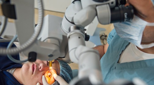
What to expect from corneal cross-linking
Keratoconus causes the cornea — the front window of the eye — to be distorted and shaped like a cone.
The abnormal shape can cause vision changes, such as light sensitivity, glare and irritation, and can lead to frequent changes in glasses and contact lens prescriptions for nearsightedness.
The cornea contains tiny fibers of protein called collagen, and these fibers help hold the cornea in place and keep it from distorting.
Corneal cross-linking involves removing the front layer of the cornea and administering an eye drop of liquid riboflavin (vitamin B12) to the surface of the eye. Ultraviolent light is then delivered to the eye at differing levels of time and intensity. The eye drop helps the cornea absorb the ultraviolet light and stiffen.
The procedure lasts 60 to 90 minutes. Patients will see results after six months to one year.