Newswise — NEW YORK, NY (Jan. 15, 2020)—The source of essential tremor—a movement disorder that causes involuntary trembling of the hands, arms, and head—has been enigmatic, impeding the development of effective treatments for a condition that affects 4% of people over 40.
Now a new study from Columbia University Irving Medical Center and NewYork-Presbyterian suggests the tremors are caused by overactive brain waves at the base of the brain, raising the possibility of treating the disorder with neuromodulation to calm the oscillations.
“Past studies have identified changes in brain structure in people with essential tremor, but we didn’t know how those changes caused tremors,” says Sheng-Han Kuo, MD, the study’s senior author and assistant professor of neurology at Columbia University Vagelos College of Physicians and Surgeons.
“This study pins down how those structural changes affect brain activity to drive tremor.”
The study was published online today in Science Translational Medicine.
About Essential Tremor
Essential tremor is the most common movement disorder in the United States, affecting about 10 million Americans (approximately eight times as many people as Parkinson’s disease). The condition causes involuntary, rhythmic trembling, usually in the hands, and is exacerbated during such activities as buttoning a shirt or using utensils. Although essential tremor is not life-threatening, it can severely impact quality of life.
Some beta blockers and anti-epileptic drugs can reduce symptoms, but they carry side effects, such as fatigue and shortness of breath. They also don’t work very well in essential tremor patients, which Kuo says isn’t surprising since the cause of the condition hasn’t been well understood.
Tremor Patients Have Excessive Brain Activity in the Cerebellum
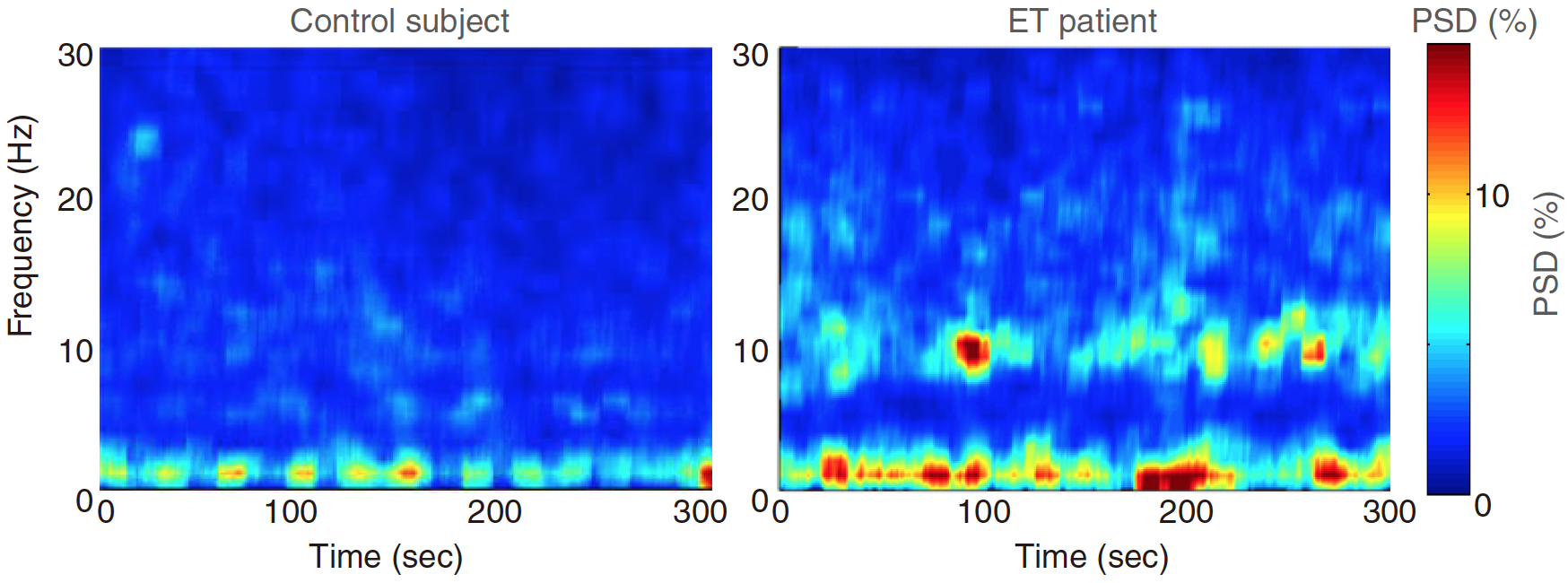
The researchers have previously identified structural changes in the cerebellum of essential tremor patients and used a new cerebellar encephalogram (EEG) technique to search for unusual brain waves in this part of the brain.
Among 20 essential tremor patients examined with cerebellar EEG, most had strong oscillations (between 4 and 12 Hz) in the cerebellum that were not found in any of the 20 control subjects. Patients with more severe tremors had stronger oscillations.
Oscillations First Found in Mice
The researchers first discovered the cerebellar oscillations in mice that had developed tremors closely resembling those seen in essential tremor patients.
The tremors could be turned on and off by stimulating certain neurons in the mouse brain, alternately suppressing and unleashing the oscillations. “These results established a causal relationship between the brain oscillations and tremor, which cannot be directly tested in patients,” says Kuo, who is also an assistant attending neurologist at NewYork-Presbyterian/Columbia University Irving Medical Center.
Excessive Oscillations Stem from Extra Synapses
In previous studies of postmortem brain tissue from essential tremor patients, the Columbia team discovered that patients with essential tremor had an abnormally high number of synapses, or connections, between two types of nerve cells in the brain’s cerebellum—climbing fibers and Purkinje cells.
In the current study, again using postmortem brain tissue, the researchers found that the formation of these synapses appears to be influenced by a protein called glutamate receptor delta 2 (GluRẟ2). “When this protein is underexpressed, any excess synapses that form between climbing fibers and Purkinje cells are not eliminated, resulting in too many neuronal connections,” says Kuo.
When the team reduced expression of GluRẟ2 in mice, the animals developed tremors similar to those seen in humans. Restoring GluRẟ2 function suppressed the tremors, proving that the protein plays a key role in essential tremor.
Potential for New Treatments
The study opens several new possibilities for treatment of essential tremor, Kuo says.
“Using cerebellar EEG as a guide, we may be able to use neuromodulation techniques such as tDCS or TMS (transcranial direct-current stimulation or transcranial magnetic stimulation) to reduce tremor, or even drugs to reduce transmission between the climbing fibers and Purkinje cells.”
Kuo is also working to develop medications that increase GluRẟ2 expression in the brain, which may reduce tremor.
+++
The study is titled “Cerebellar oscillations driven by synaptic pruning deficits of cerebellar climbing fibers contribute to tremor pathophysiology.”
The other contributors are Ming-Kai Pan (National Taiwan University Hospital, Taipei City, Taiwan), Yong-Shi Li (Columbia University Irving Medical Center, New York, NY), Shi-Bing Wong (CUIMC and Taipei Tzu Chi Hospital, Tzu Chi Medical Foundation, New Taipei City, Taiwan), Chun-Lun Ni (CUIMC), Yi-Mei Wang (National Taiwan University Hospital), Wen-Chuan (National Taiwan University Hospital), Liang-Yin Lu (National Taiwan University Hospital), Jye-Chang Lee (National Taiwan University Hospital), Etty P. Cortes (CUIMC), Jean-Paul G. Vonsatte (CUIMC), Qian Sun (Columbia and Case Western Reserve University, Cleveland, OH), Elan D. Louis (Yale University, New Haven, CT), and Phyllis L. Faust (CUIMC).
The research was supported by the National Institutes of Health (grants K08NS083738, R01NS104423, R01NS086736, R01NS073872, R01NS085136, R01NS088257, R01NS04289, and R21NS077094), Parkinson’s Foundation, International Essential Tremor Foundation, National Institute of Environmental Health Sciences, Ministry of Science and Technology in Taiwan, and National Taiwan University Hospital, and a Louis V. Gerstner Jr. Scholar Award.
The authors declare that they have no financial or other conflicts of interest.